
Published: 07/02/2023|Last Updated: 01/01/2024
In Biology, specifically in Microbiology Gram Staining is an inevitable technique. It was introduced in the year 1884 by Hans Christian Gram. This staining process can classify a bacteria into Gram Positive and Gram Negative. It can be used as a step in various bacterial diagnosis.
|
Table of Contents
|
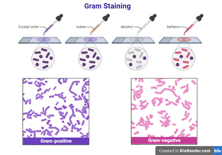
Reagents Used
- Primary stain – Crystal Violet
- Secondary stain or counter stain – Safranin
- Gram’s Iodine (Mordant – Used along with Crystal Violet)
- Acetone and Alcohol based decolorizing agent.
Principle
It is the process in which the bacteria is stained with a series of reagents used for gram staining. It is initially stained with the primary stain which is Crystal Violet. The primary stain is fixed by iodine. Then it is treated with alcohol so that decolourization will take place. Some bacteria can retain the stain but some cannot. So the bacteria which retains the Crystal Violet – Iodine mixture is called Gram-Positive Bacteria. They will appear in blue or purple color. The retaining of the stain complex is due to the cell wall of the bacteria. During decolorization, the pores in the cell wall containing the stain complex dehydrate and shrink. So the stain cannot move out and decolorization will not take place.
The cell wall of the Gram-Negative bacteria is very thin. It cannot retain the Crystal Violet – Iodine mixture. So during destaining, the stain mixture will be washed off. For identification, it is again stained with safranin. This bacteria take up the safranin and look red/pink in color.
Procedure


- Take a freshly cleaned glass slide.
- Smear the sample into the slide with the appropriate smearing procedure.
- Dry the sample at room temperature and then gently heat-fix it.
- The primary dye Crystal Violet must be poured over the slide containing the sample and kept for about 60 seconds and rinsed with water.
- Add the mordant (Gram’s Iodine), keep it for 60 seconds, and wash it with water.
- Add 95% alcohol or acetone or a mixture of both used for rapid decolorization.
- Add safranin for 60 seconds (Counter staining).
- Dry the slide and observe under the microscope.
JobFinder Listing
Result
 |
- Gram Negative: Red/Pink Color
- Gram Positive: Blue Color
Purpose
Gram staining is used to identify the bacteria which cause certain infections. This can be done using blood, body fluids, etc. This helps to give clear information to start medication. This is not the final or only way of diagnosis. This is only used to classify the bacteria. All the bacteria cannot be classified by this method.
Gram-Positive Bacteria
It is a type of bacteria which have a thick peptidoglycan outer cell wall. This peptidoglycan layer causes Gram-Positive bacteria to look Blue by Gram Staining.
Examples: Streptococcus species, Clostridium species, etc.
Gram-Negative Bacteria
It is a type of bacteria having thin layers of a peptidoglycan layer. This causes the Gram stain to get washed away and retains the counter stain safranin to retain and appear Red.
Examples: E.coli, Pseudomonas species, etc.
Download this Note as PDF
Image Source: Created with BioRender.com
Published: 08/02/23, 0053