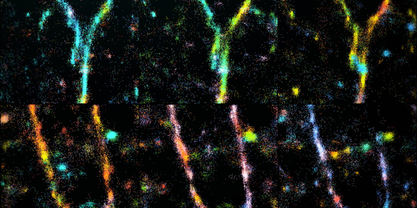
Researchers can now discern orientation and other minute characteristics in the nanostructures of biological systems that were previously unseen thanks to a new imaging technology developed by the Lew lab.
Microscopy
By combining two microscopy techniques single-particle analysis and cryo-electron tomography, or cryo-ET researchers created a unique imaging method for visualizing mitochondria from animal models.
The electron microscope was developed during the 1930s by Ernst Ruska and was first applied to biological specimens by Albert Claude, Keith Porter, and George Palade. The use of electron beams to deflect and focus that beam using electrostatic and magnetic coils or lenses is the basic principle of an electron microscope.
Microscopy is a vital tool used in scientific research and analysis. It involves the use of specialized instruments to observe and study microscopic objects or structures. There are different types of microscopes available, but two of the most commonly used are light microscopy and electron microscopy. In this article, we […]
A microscope is a device used to visualize materials with a micro size that is not visible to the naked eye. The first microscope was invented by Zacharias Janssen. This type of microscope is called the light microscope as it uses visible light to illuminate the object. The light […]