Prostate cancer is the second leading cause of cancer-related fatalities in the United States, making it a persistent danger to the health of men. In the United States, over 250,000 men are diagnosed with prostate cancer annually. Although the majority of cases have modest rates of morbidity and death, a small percentage of patients require intensive care. The primary tool used by urologists to determine if such therapy is necessary is the Gleason score, which rates the appearance of the prostate gland on histology slides. There is a great deal of variation in interpretation, though, which can result in both overtreatment and undertreatment.
There are drawbacks to the existing approach, which uses histology slides. Not all of the biopsy is examined in two dimensions (2D), which increases the chance of missing important features. Additionally, complex 3D glandular systems may be difficult to comprehend when viewed on 2D tissue slices. Furthermore, tissue is destroyed during standard histology, which restricts further studies. To overcome these drawbacks, scientists have created nondestructive 3D pathology techniques that provide full biopsy specimen imaging while maintaining tissue integrity.
There are drawbacks to the existing approach, which uses histology slides. Not all of the biopsy is examined in two dimensions (2D), which increases the chance of missing important features. Additionally, complex 3D glandular systems may be difficult to comprehend when viewed on 2D tissue slices. Furthermore, tissue is destroyed during standard histology, which restricts further studies. To overcome these drawbacks, scientists have created nondestructive 3D pathology techniques that provide full biopsy specimen imaging while maintaining tissue integrity.
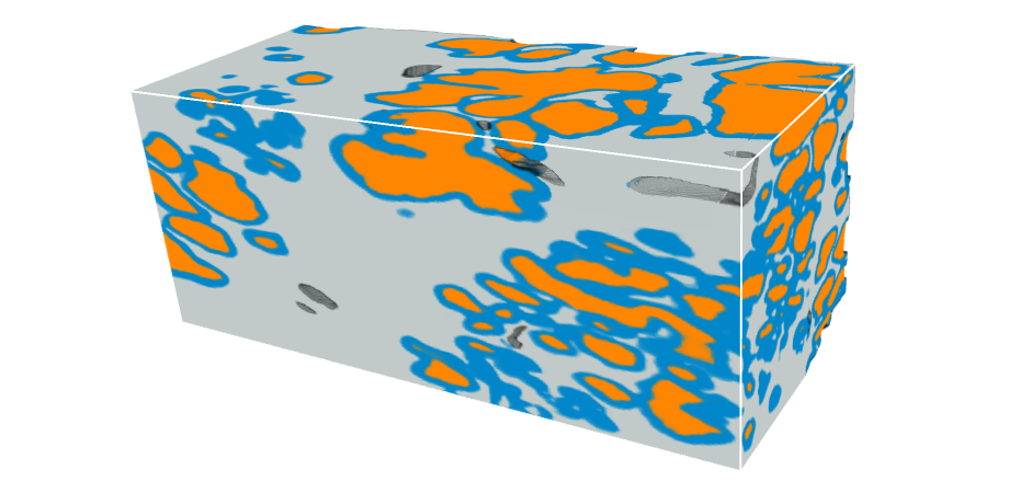
Methods for acquiring 3D pathology information have been developed recently, improving prostate cancer risk assessment. By creating a deep learning model, research published in the Journal of Biomedical Optics (JBO) fully utilises the potential of 3D pathology to enhance the 3D segmentation of glandular tissue structures, which are essential for assessing the risk of prostate cancer.
Under the direction of Professor Jonathan T. C. Liu of the University of Washington in Seattle, the study team trained nnU-Net, a deep learning model, directly using 3D prostate gland segmentation data that was acquired via earlier intricate procedures. Their group’s open-top light-sheet (OTLS) microscopes were used to get the 3D datasets of prostate biopsies, from which their model effectively produces 3D semantic segmentation of the glands. Understanding the tissue composition is essential for prognostic studies, and the 3D gland segmentation offers insightful information on this front.
Our results indicate nnU-Net’s remarkable accuracy for 3D segmentation of prostate glands even with limited training data, offering a simpler and faster alternative to our previous 3D gland-segmentation methods. Notably, it maintains good performance with lower-resolution inputs, potentially reducing resource requirements.
Jonathan T. C. Liu
The novel 3D segmentation model for prostate cancer, which is based on deep learning, is a major advancement in computational pathology. It has the potential to guide important therapeutic decisions and eventually improve patient outcomes by enabling precise characterisation of glandular structures. This development emphasises how computational methods might improve medical diagnosis. It has the potential to advance personalised medicine in the future by opening the door to more focused and successful therapies.
Also Read| Micro- and nanoplastics in the body are passed on during cell division
Beyond the constraints of traditional histology, computational 3D pathology provides the capacity to get important insights into the course of illness and customise therapies to meet the specific requirements of each patient. In the fight against prostate cancer, new frontiers of accuracy and potential are being opened by researchers who are persistently pushing the envelope of medical innovation.
Source: The International Society for optics and photonics – News
Journal Reference: Wang, Rui, et al. “Direct three-dimensional segmentation of prostate glands with nnU-Net.” Journal of Biomedical Optics 29.3 (2024): https://doi.org/10.1117/1.JBO.29.3.036001
Last Update:


