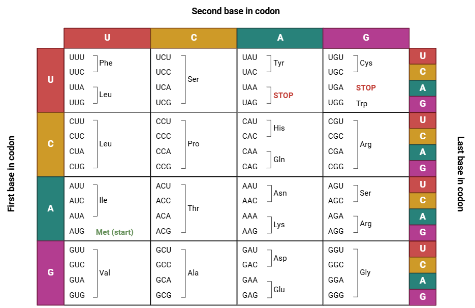
The codon chart is an important tool in the fields of molecular biology and genetics, allowing the translation of the genetic code into specific amino acids, creating the basis for protein synthesis in all life forms. The interpretation of the codon chart implies comprehension of the structure of genetic material (DNA and RNA) as well as the mechanisms of transcription and translation and especially the mechanisms of how codons determine what amino acid sequence follows.
Interpreting a Codon Chart
A codon chart visually represents the relationship between codons and amino acids. When using it, locate the three nucleotides of a codon by position. The chart helps identify which amino acid corresponds to each codon in mRNA.
Codon Chart Layout

There are different types of codon charts, but the two most usual kinds are:
The Table Format: The codon chart is arranged systematically, usually in tabular form, mainly as rows and columns, such that each individual cell represents a particular codon, along with the amino acid it encodes.
The Wheel Format: The circular codon chart allows the determination of the corresponding amino acid by starting from the center where the first base of the codon lies and then extending outwards to include the second and the third bases.
Also Read: Amino Acids – Properties, Structure and Classes
How to Use a Codon Chart to Pinpoint Your Amino Acids?
Start by identifying the first letter of the codon and locate the corresponding nucleotide on the table. Now advance to the second letter of the codon and determine what the column or segment. Finally, the third letter is determined to identify the proper amino acid or termination signal.
For example: Codon UUU: Selecting U in the first position, followed by U in the second and U in the third position, allows one to get that UUU is associated with the amino acid phenylalanine. The AUG codon is designated as the initiation codon and codes for the amino acid methionine while also initiating translation.
Decoding the 64 Codons
There are 64 possible codons, all the permutations of the four RNA bases in triplets. The coding includes:
61 Codons for Amino Acids: Since there exist only 20 amino acids, most of the amino acids are represented by more than one codon. Thus, the genetic code is redundant or degenerate.
3 Stop Codons: UAA, UAG, and UGA are the stop signals that define the termination of a protein-coding sequence, stopping translation.
Amino Acid Redundancy
The redundancy inherent in the genetic code enables certain amino acids to be represented by several codons, a characteristic that confers robustness against genetic mutations. For instance:
- Serine is coded by six different codons: UCU, UCC, UCA, UCG, AGU, and AGC.
- Leucine is coded by six different codons: UUA, UUG, CUU, CUC, CUA, and CUG.
Because of this degeneracy, even though the mutation modifies one nucleotide, the same amino acid codon can still be expressed, although with a diminished impact on the protein’s functionality.
Also Read: Meiosis: Definition, Stages, Mechanism, and Diagram
Start and Stop Codons
Start Codons
The start codon AUG, thus serves a dual purpose. It indicates the beginning translation of the mRNA.
It codes for methionine, an essential amino acid for the initiation of protein synthesis.
Stop Codons
The stop codons, UAA, UAG, and UGA, signify no amino acid code. They are the end of the translation. The moment a ribosome meets one such codon, it expels the polypeptide chain that has been synthesized.
Also Read| Glycolysis – Pathway and Process Overview
Analyzing genetic mutations using a codon chart
Silent Mutations: Nucleotide sequence change which doesn’t alter the amino acid concerned because of redundancy in genetic code.
Missense Mutations: Changes that result in a different amino acid, potentially altering the protein’s function.
Nonsense mutations: Changes that convert an amino-acid-encoding codon into a stop codon, truncating the protein.
Codon Wheel

Structure of codon wheel
Inner Circle: The innermost circle of the wheel defines the first base, the first base or nucleotide, in the RNA codon sequence. It can be one of the four variants: Adenine (A), Uracil (U), Cytosine (C) or Guanine (G).
Middle Ring: Continuing outward, the second ring represents the second base of the codon. This, it turns out, is actually the base found through extension from the first base out toward the strand being extended.
Outer Circle: The third base or final nucleotide that separates one codon from another is represented by this circle. It chooses which particular amino acid will be encoded.
Amino Acids: Positioned along the rim of the wheel, these amino acids are listed with their codons. Each three-nucleotide RNA codon is translated into an abbreviation for an amino acid-for example, “Phe” is phenylalanine-or a stop signal.
Image Source: BioRender
Last Modified:
Graduated from the University of Kerala with B.Sc. Botany and Biotechnology. Attained Post-Graduation in Biotechnology from the Kerala University of Fisheries and Ocean Science (KUFOS) with the third rank. Conducted various seminars and attended major Science conferences. Done 6 months of internship in ICMR – National Institute of Nutrition, Hyderabad. 5 years of tutoring experience.


