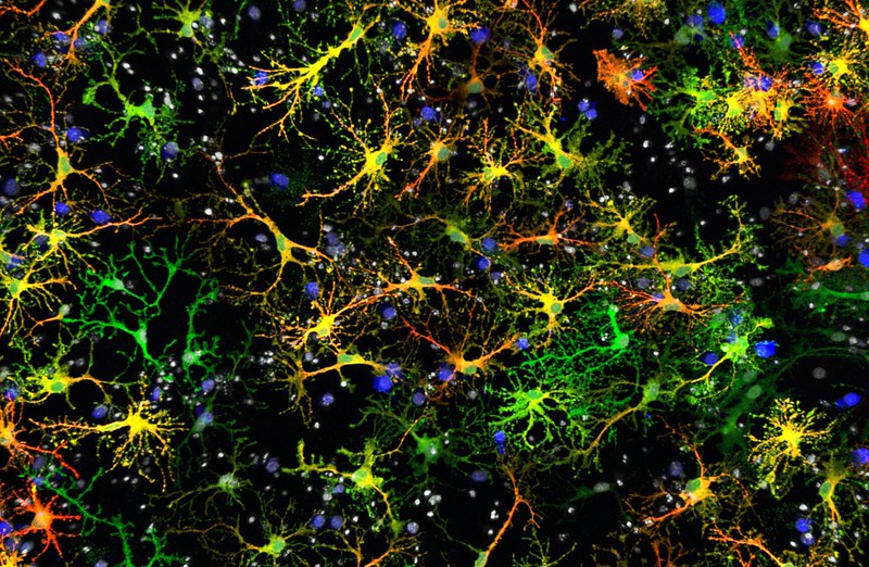
Cellular changes that herald the early stages of Alzheimer’s disease have been catalogued in unprecedented detail for the first time in more than 1.6 million brain cells from elderly individuals in new routes by which to seek ways to prevent the most prevalent cause of dementia among older people.
Their findings were published in the journal Nature.
It also recognized a separate community of cells driving the older brain down another route that does not seem to lead to Alzheimer’s disease.
Our study highlights that Alzheimer’s is a disease of many cells and their interactions, not just a single type of dysfunctional cell,
We may need to modify cellular communities to preserve cognitive function, and our study reveals points along the sequence of events leading to Alzheimer’s where we may be able to intervene.
Philip De Jager
It was a technical tour de force, new molecular technologies, machine-learning techniques, and a large collection of brains donated by aging adults in an ingenious way.
While these earlier studies of brain samples from Alzheimer’s patients have given clues to the molecules involved, they haven’t revealed much about where those genes in turn act within the long sequence of events that leads to Alzheimer’s which cells are involved in each step of the process.
Past studies have analyzed brain samples as a whole and they lose all cellular detail,
We now have tools to look at the brain in finer resolution, at the level of individual cells. When we couple this with detailed information on the cognitive state of brain donors before death, we can reconstruct trajectories of brain aging from the earliest stages of the disease.
Philip De Jager
The new analysis required more than 400 brains, courtesy of the Religious Orders Study and the Memory & Aging Project based at Rush University in Chicago.
The researchers collected several thousand cells in each brain from a region of the brain that is affected by Alzheimer’s and ageing. They then ran each cell through a process called single-cell RNA sequencing, which gives a readout of the cell’s activity and which of its genes were active.
Next, algorithms and machine-learning techniques, developed by Menon and Habib, analyzed data from all 1.6 million cells to identify the types of cells present in the sample, including how different cells interacted with one another.
These methods allowed us to gain new insights into potential sequences of molecular events that result in altered brain function and cognitive impairment,
This was only possible thanks to the large number of brain donors and cells the team was fortunate enough to generate data from.
Vilas Menon
These brains were able to resolve one of the major hurdles in research on Alzheimer’s: determining the order in which cellular changes occur in Alzheimer’s and distinguishing these changes from those typical of normal ageing of the brain, as the brains were from persons at many stages of the disease process.
We propose that two different types of microglial cells—the immune cells of the brain—begin the process of amyloid and tau accumulation that define Alzheimer’s disease.
Philip De Jager
Following pathology accrual, the cells involve another cell type astrocytes that play an important role in the alteration of electrical connectivity in the brain, resulting in cognitive impairment. Interacting with each other the cells bring additional cell types resulting in serious disruptions in the functioning of the human brain.
These are exciting new insights that can guide innovative therapeutic development for Alzheimer’s and brain aging,
By understanding how individual cells contribute to the different stages of the disease, we will know the best approach with which to reduce the activity of the pathogenic cellular communities in each individual, returning brain cells to their healthy state.
Philip De Jager
Source: Columbia University Irving Medical Center – News
Journal Reference: Green, Gilad S., et al. “Cellular Communities Reveal Trajectories of Brain Ageing and Alzheimer’S Disease.” Nature, 2024, pp. 1-12, https://doi.org/10.1038/s41586-024-07871-6.
Last Modified:


