
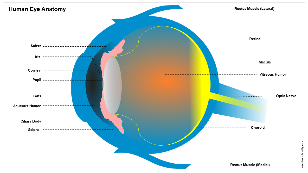
The eyes are a major organ in Humans similar to every other organism. It has a highly complex structural orientation which produces a sense of vision. It has complex receptors of light that can gather the surroundings and convert them into electrical signals/nerve impulses, which the brain processes and interprets to produce vision. Humans have binocular vision. It is the method by which both eyes can produce a single enlarged and wide image. A detailed labelled human eye diagram is attached above.
The working of the eye is highly complicated. It undergoes several muscular adjustments that allow and restrict the entry of light into the inner parts which control the various vision formation. This is a continuous process as the sense of vision is produced.
Three Layered Structure
Human eyes are a triple-layered structure. More deeply it can be explained in three parts
- External coat (fibrous)
- Middle coat (vascular)
- Internal Coat (Nervous)
External coat (fibrous)
The external fibrous coat is the outermost layer of the eyes. This layer is composed of the cornea and sclera. The cornea covers 1/6 of the total eye. The sclera plays a major role in the physical activity of the eyes which supports muscular movement.
Middle coat (vascular)
This is a median between the anterior and posterior parts of the eyes. This layered is composed of ciliary muscle fibres, iris, and choroid. The choroid is composed of a dark pigment called melanin which helps in the reflection of eyes. Iris acts similarly to the camera literature of the camera covers over the lens. Ciliary fibres play a major role in the physical movement of the structure which helps in the alteration of the lens shape for focusing the object.
Internal Coat (Nervous)
The innermost layer of the eyes is the retina which is a key player in vision. It consists of the photoreceptors and the nerve cells transmitting the impulses. The major photoreceptors in the inner layer/retina are the rods and cones. Cones undergoes the reception of the colours in daylight. Rods support black & white vision.
Read more| Good Laboratory Practice (GLP) – Overview [PDF]
Parts of an Eye
An eye is composed of many complex parts that work in sync to produce vision. They can be classified as:
Cornea
The clear front portion of the eye covers the pupil and iris. It facilitates directing inbound light onto the lens.
Iris
The pigmented portion of the eye is called as the iris. It covers the centremost opening of the eyes called as the pupil. This acts as a regulator to control the intensity of the light that leads to the inner portion of the eye.
Pupil
This is the centermost opening in the iris with a dark shade. With the muscular action, it can vary its size to alter the amount of light passing. Due to this mechanism of the pupil, we may feel some short-term darkness in our vision when moving from an area with a brighter light to an area in minor darkness.
Lens
The lens of the human eye is a flexible structure, that can alter its shape and size with the action of the ciliary muscles. This mechanism makes the lens to focus the object at varied distances on the retina.
Ciliary Muscles
These are the muscular fibres that help in the physical movement within the lens which helps in the focusing of near and away objects.
Retina
The retina is the innermost layer of the eye. On the retina, the lens focuses the light from the object and forms images. The layer of the retina is composed of the light receptors or the photoreceptor cells rods and cones. These cells can capture the incoming light and then it can be converted to electric impulses. These impulses are transmitted to the brain through optic nerves for interpretation.
Photoreceptors
- Rods: This photoreceptor is responsible for the vision in low-light conditions.
- Cones: These photoreceptors are responsible for coloured vision.
Read more| Simple Microscope – Definition, Structure and Applications
Aqueous Humor
Aqueous humor is a transparent liquid that plays a key role in the filling of the empty portion in the front portion of the eye, near the lens and cornea.
Vitreous Humor
This is a transparent fluid or a gel-like structure that fills the space between the lens and the retina provides support for it and maintains the shape of the eye.
Optic Nerve
A collection of nerve fibres arises from the retina to the brain transmitting the nerve impulses after the conversion of the collected light from the object by the photoreceptors.
Sclera
The part that provides physical support to the eyes and its protection.
Choroid
It is the layer filled between the retina and sclera which contains the blood vessels that support circulation.
Conjunctiva
The outermost layer of the eyes behind the eyelids prevents any direct damage to the eyes. It is the layer affected by the disease conjunctivitis.
Author: Neethu Krishna | Edited: Ajmal Aseem
Last updated:
Also Read


![Good Laboratory Practice (GLP) - Overview [PDF]](https://cdn.biotechreality.com/wp-content/uploads/2023/10/GLP_Cover-min-300x169.png)