
The process of replication of chromosomes and then its segregation by a cell producing two identical daughter cells is called as the mitotic cycle. The M phase of the cell cycle comprises this mitosis along with cytokinesis.
Four major steps are involved in Mitosis which are,
- Prophase
- Metaphase
- Anaphase
- Telophase
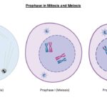
Prophase
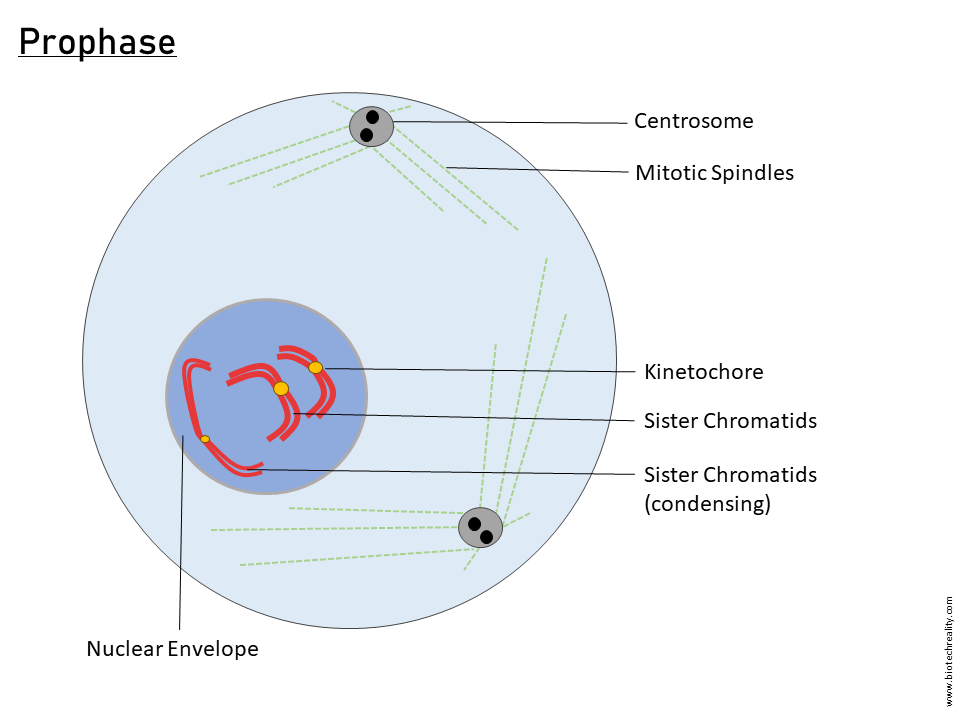
The prophase can be defined as the early duration of a mitosis which involves various events that are essential for the other steps.
Such events include,
- The disassembling of the nuclear membrane or the membrane retracts back to the Endoplasmic reticulum – the structural integrity of the nuclear membrane is mainly provided by lamins. The degradation of lamins leads to the retraction of the nuclear membrane.
Read More: Escherichia coli or E. coli – Structure and Function
Degradation of lamins
The M phase Cyclin-dependent kinase 1 (CDK 1) phosphorylates lamins. Lamin A and C disassociate as soluble subunits after the phosphorylation and Lamin B becomes a subunit of membrane vesicles. The cdk1 also phosphorylates ER, Golgi proteins etc and disassembles them.
- Nucleolus disassembles
- Chromatin higher-order condensation begins – In the early stage of prophase, the condensation starts with 30 nm and then becomes 300nm and when the nuclear membrane retracts it condenses to 700nm chromatids.
- The internal membrane system disassembles to double the entire cell contents for equal distribution
- The membrane receptors and markers get internalized
- Endocytosis and exocytosis are halted
- Cells become round
- The interface array of microfilaments and microtubules is reorganised
- Kinetochore get assembled into the centromere of chromosomes
Kinetochore attachment
It is a multiprotein complex consisting of an inner plate, outer plate and fibrous corona. The kinetochore attaches to the heterochromatic centromere which also contains some proteins.
Proteins seen in,
Centromere: INCENP, CENP A, CENP B, CENP G etc.
Inner Plate: CENP A, CENP C, CENP G etc.
Outer plate: CENP E, CENP F, CENP I, one of the AP2A family proteins, NDC 80 complex etc.
Fibrous corona: Mad1, Mad2, Bub 1, Dam 1 complex etc.
Later in the prophase, the chromatin becomes chromosomes. For further steps, the microtubule directionality is provided by the Ran protein gradient. Ran is a monomeric G protein; the microtubules polymerize towards the direction where the Ran GTP is higher.
The GEF or Guanine nucleotide exchange factor of Ran is present in the nucleus as well in the kinetochore and its GAP or GTPase activating protein is present in the cell membrane or cell cortex that is towards the cytoplasmic side. After nuclear membrane disassembly, the polymerization of microtubules to the Ran GTP region in the kinetochore takes place and the microtubules get attached to the kinetochore and establish the kinetochore microtubules.
The microtubule that passes the kinetochore without attaching them i.e., passing from one pole to the other are the inter-polar microtubules. From here the Prometaphase starts.
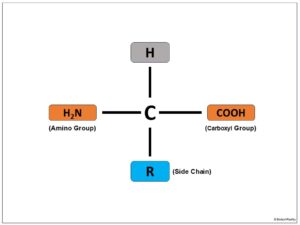
Read More: 20 Types of Amino Acids – Details and Structure
Prometaphase

The phase just before the metaphase of mitosis is required for the proper establishment of metaphase. For the alignment of the chromosome at the metaphase plate microtubules stretch from the kinetochore.
The attachments are of different types,
- Monotelic Attachment – microtubule from one pole to the kinetochore but there will be no stretching force to separate the outer plate away from the inner plate of the kinetochore.
- Merotelic Attachment – microtubule from opposite pole attaches to the same kinetochore, hence there will be no stretch.
- Syntelic Attachment – microtubule from one pole attaches to kinetochores on two sides and hence no stretch.
- Amphitelic Attachment – microtubules from opposite poles attach to opposite kinetochores, so the chromosomes move but do not spit as there is cohesion.
From these types, the amphitelic attachments are retained and others are avoided.
The protein present in the Fibrous corona of the kinetochore NDC80 attaches with the microtubule in its non-phosphorylated form and it is done by the outer plate protein PP2A (protein phosphatase 2A), but when the outer and inner plates are attached the aurora B kinase in the inner kinetochore phosphorylates the NDC80. When amphitelic attachment occurs, as the outer plate stretches away from the inner kinetochore, the aurora B kinase does not phosphorylate the NDC80 and hence the attachment of the microtubule with the kinetochore becomes stabilized.
Read More: Meiosis: Definition, Stages, Mechanism, and Diagram
Metaphase

Here starts the metaphase as the chromosome senses equal force generated from poles and the alignment of the chromosome at the metaphase plate takes place. The process by which a chromosome moves to the metaphase plate is called chromosome congression. For the chromosome congression to take place the short microtubules have to grow and the long has to shrink, Kinesin 7 protein gets attached to the short microtubule side of the kinetochore and leads to the polymerization of short microtubules and kinesin 13 depolymerizes the long microtubule side of the kinetochore. The dynein-dynactin complex binds both kinetochores of chromosomes favours chromosome congression. As these forces are concentrated on the centromere kinesin 4 or chromokinesin for movement of the chromosome arm. When all chromosomes align at the metaphase plate, the metaphase is established.
Metaphase to anaphase transition
It is controlled by the spindle assembly checkpoint which is already initiated from prophase during the kinetochore assembly. During the kinetochore assembly, the proteins Knl1 in fibrous corona get phosphorylated by MPS1 kinase and knl 1 phosphorylates the other two proteins bub3, bub 1 kinase, and bubR1. These phosphorylated proteins activated mad 1 and then mad 2 now mad 2 comes to its active closed conformation. This protein complex now works as a Mad 2 activating centre, so every mad 2-o (open) becomes a mad 2-c(closed) active form when they interact with the complex. Within the prophase all mad 2 become active and they form another complex called MCC, a mitotic checkpoint complex that binds and interacts with APC/Ccdc20 (Anaphase promoting factor) for the establishment of Anaphase.
Anaphase

The stage is where the separation and segregation of sister chromatids to opposite poles. During the transition from metaphase to anaphase the chromosome condensation protein called cohesin has to be cleaved which is done by the seprase that cleaves the Kleisin family proteins and the transition completes and anaphase begins.
Anaphases A and B are there, anaphase A involves the movement of sister chromatids towards the poles and anaphase B involves the movement of centrosomes to extreme poles.
Anaphase to Telophase transition
It is regulated by spindle position checkpoint and this was studied in the budding yeast. The different proteins shown in the image have different functions also, LTE 1 ensures the kin4 is not entering into the bud cell, and Tem 1 binds with the centrosome or spindle pole body (SPB) in GDP-bound inactive form. When the proper segregation occurs during the anaphase one of the SPB enters the bud cell where kin4 is absent so Tem 1 is converted to Tem1 GTP form and becomes active, the active Tem 1 dissociates cdc14 from the nucleolus and now cdc14 is free and active. The cdec14 is the master phosphatase that regulates the mitotic exit. The cdc14 can reverse all the phosphorylation given by cdk1 and the exact reversal of prophase happens.
Read More: Osmosis – Definition, Diagram, Applications and FAQ
Telophase

The reversal of Prophase is referred to as the telophase. Nuclear reformation occurs, an internal membrane system forms and vesicles pinch off from the membrane engulfing the chromosome. These vesicles fuse and thus an inner membrane is formed, the ER extends near to this inner membrane forming an outer membrane where nuclear lamins form a structural network and other proteins attach to it and the reformation of the nucleus takes place. After telophase cytokinesis (cell division) takes place and two daughter cells are formed by mitosis.
Cytokinesis

A eukaryotic cell splits into two daughter cells, each with its own nucleus, during a process known as cytokinesis. It is essential for the growth, development, and maintenance of multicellular organisms and is the last phase of the cell cycle, coming after mitosis or meiosis. By preserving the correct number of chromosomes in each daughter cell, cytokinesis makes sure that the genetic material from the parent cell is appropriately distributed across the daughter cells.
Author: Neethu Krishna
Diagrams: Achuth B S
Last updated: