In species capable of sexual reproduction, a form of cell division known as meiosis takes place. Gametes, which are specialized reproductive cells (sperm and eggs) with half the number of chromosomes as the parent cell, are formed by a carefully controlled process.
Making sure that the developing zygote has the appropriate number of chromosomes when gametes unite during fertilization is the main purpose of meiosis. Meiosis does this by halving the number of chromosomes, guaranteeing genetic variety in progeny.
Read More: Endoplasmic Reticulum: Architecture, Function, and Structure
Meiosis is a two-step process that consists of prophase, metaphase, anaphase, and telophase phases in each division, known as meiosis I and meiosis II. The pairing of homologous chromosomes during prophase I, the crossing over (exchange of genetic material) between homologous chromosomes, and the random sorting of chromosomes during metaphase I are the main characteristics that set meiosis apart from mitosis (another kind of cell division).
Stages of meiosis and its mechanism
What are the cycles of meiosis?
Meiosis I and Meiosis II are the two consecutive cycles of cell division that makeup meiosis. Meiosis produces four genetically separate haploid cells by halving the number of chromosomes, in contrast to mitosis, which yields two identical daughter cells.
Also Read: Codon Chart and Codon Wheel – Explained
Merck’s Enpatoran FDA Breakthrough Therapy Designation Granted for Cutaneous Lupus Treatment
Merck has secured an FDA Breakthrough Therapy designation for enpatoran, an investigational oral TLR7/8 inhibitor aiming to address the high unmet need in lupus patients […]


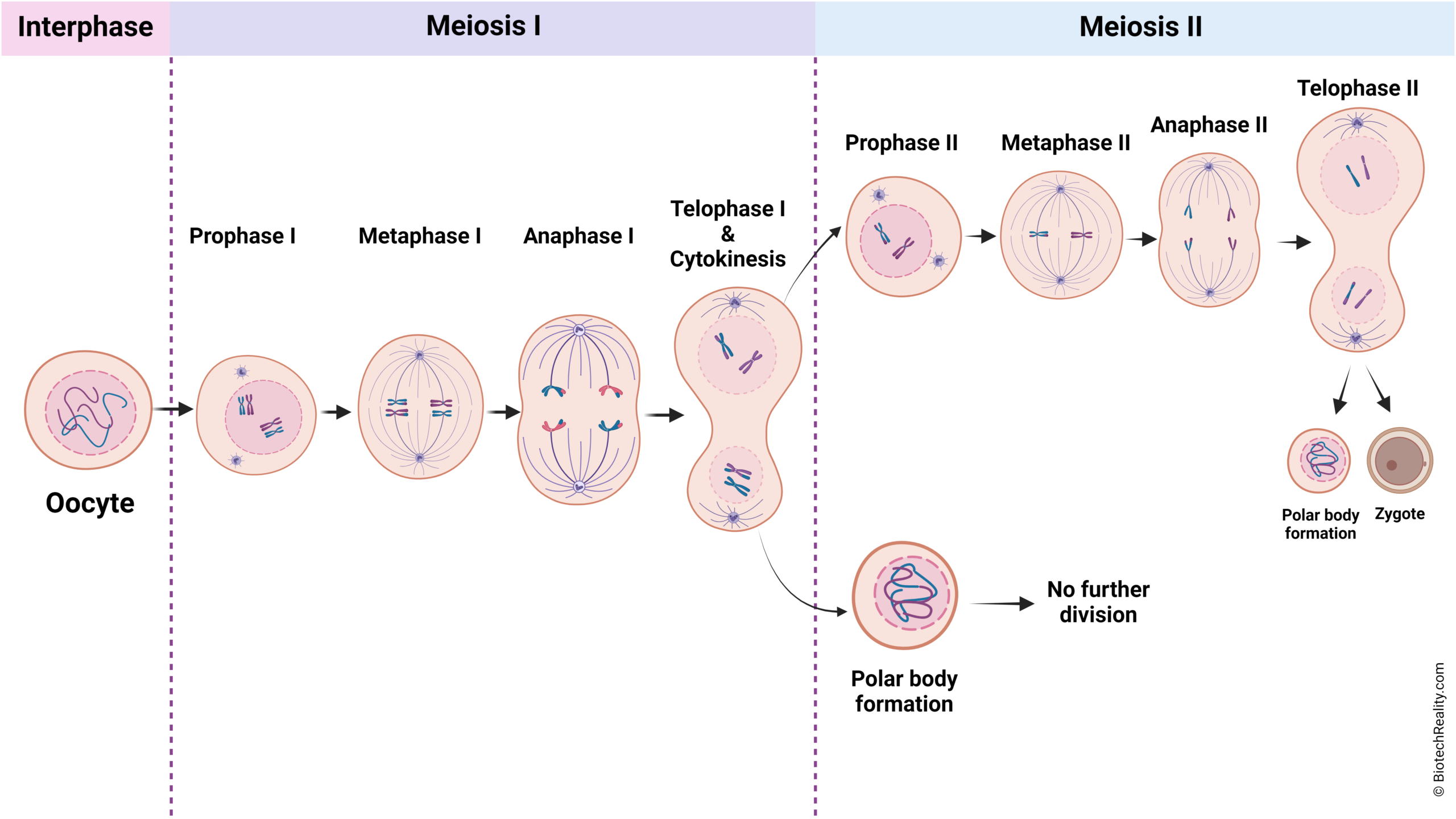
Meiosis I
Prophase I
Prophase I, the longest and most intricate step, marks the beginning of meiosis I. Chromosomes condense during this stage and become visible under a microscope. Remarkably, homologous chromosomes join together via a process called synapsis to create structures known as tetrads. Genetic diversity is increased within tetrads by crossing over, which is the exchange of genetic material between homologous chromosomes. Chiasmata arise as a result of successful crossover occurrences.
Metaphase I
The first cycle of meiosis, known as metaphase I, is the second step of the process. The homologous chromosomal pairs align near the cell’s equator during metaphase I. Because each pair may align differently due to the random nature of this alignment, the subsequent daughter cells may have a diverse genetic makeup. The homologous chromosomal centromeres are where the spindle fibres link to them, and the spindle fiber microtubules pull the chromosomes in the direction of the cell’s equator. Because it dictates which chromosomes will be split during anaphase I, the orientation of the chromosomes at the cell’s equator is significant. Because it dictates which chromosomes will be passed on to the daughter cells, chromosomal orientation is also significant for genetic variety. The cell is prepared to go on to anaphase I after the chromosomes are aligned at the equator.
Anaphase I
The third stage of meiosis I is called anaphase I. The homologous chromosomes, which are each made up of two chromatids, are dragged towards the opposing poles of the cell by the spindle fibers during anaphase I. The homologous chromosomes separate when the spindle fibers shorten and compress. During anaphase I, the sister chromatids of every chromosome stay together at the centromere and do not split. To ensure that each daughter cell receives a distinct mix of chromosomes, resulting in genetic diversity, the separation of homologous chromosomes during anaphase I is crucial. The cell is prepared for telophase I once the homologous chromosomes have split apart.
Telophase I and Cytokinesis
The first cycle of cell division in meiosis, known as telophase I, is the fifth step of the process. The spindle fibres vanish and the paired homologous chromosomes go to the cell’s poles during telophase I. Each pair of chromosomes causes the nuclear envelope to reorganise, isolating the nuclear DNA from the cytoplasm. The chromosomes become less compact and more dispersed as they start to uncoil. The process of cytokinesis, which separates the parental cell’s cytoplasm into two daughter cells, occurs in conjunction with telophase I in the life cycle of the cell. Half as many chromosomes as the parent cell, the daughter cells that develop are haploid. The next stage might be an interkinesis, depending on the species (a short stage before the second meiotic division) or the daughter cells might go straight into meiosis II prophase II. After meiosis I, daughter cells are created that have distinct chromosomal makeup.
The process of splitting the parent cell’s cytoplasm into two daughter cells is known as cytokinesis in meiosis I. Following telophase I, the fifth step of meiosis I, is this procedure. The spindle fibres vanish and the paired homologous chromosomes go to the cell’s poles during telophase I. Each pair of chromosomes causes the nuclear envelope to reorganise, isolating the nuclear DNA from the cytoplasm. The chromosomes become less compact and more dispersed as they start to uncoil. The process of cytokinesis, which separates the parental cell’s cytoplasm into two daughter cells, occurs in conjunction with telophase I in the life cycle of the cell. Half as many chromosomes as the parent cell, the daughter cells that develop are haploid.
Read More: Mitosis: Definition, Stages, Mechanism of Cell Division, and Diagrams
Meiosis II
Prophase II
Meiosis II, the second wave of cell division in meiosis, includes prophase II. The nuclear envelope breaks down, the centrosomes move to the opposing poles of the cell, and the chromatin condenses into chromosomes during prophase II. To set up sister chromatid separation in the next division, the spindle machinery is rebuilt. Prophase II does not include DNA replication, in contrast to prophase I. Prophase II is less complex than prophase I and is more like the somatic cell division process mitotic prophase. It entails breaking down the nuclear membrane, chromatin condensation into chromatids and subsequently chromosomes, and getting ready for the division that will produce four haploid cells. Prophase I and prophase II are primarily distinguished by their complexity and number of steps.
Read More: 20 Types of Amino Acids – Details and Structure
Metaphase II
After prophase II, metaphase II is the second step of meiosis II. The metaphase plate, a hypothetical flat surface that divides the cell in half, is where the chromosomes align during metaphase II. Unlike in metaphase I, when the chromosomes appeared in homologous pairs, at this stage they are now distinguishable separately. Spindle fibres, which connect to the kinetochores at the centromeres of the chromosomes, hold the chromosomes in place. Between prophase II and anaphase II, or metaphase II, the sister chromatids of each chromosome split and go to the opposite poles of the cell.
Anaphase II
The phase of meiosis II known as anaphase II occurs when each chromosome’s sister chromatid splits off and travels to the opposing ends of the cell. The sister chromatids are pulled apart by the kinetochore microtubules, which shrink and aid in this separation. After metaphase II, when the chromosomes align at the cell’s equatorial plane, comes anaphase II. For each daughter cell to get a full complement of chromosomes, sister chromatids must travel to opposing poles during anaphase II. Comparable to anaphase in mitosis, anaphase II also involves sister chromatids being separated and moving in different directions towards the poles of the cell.
Read More: Amino Acids – Properties, Structure and Classes
Telophase II and Cytokinesis
The last phase of meiosis II, or the second cycle of cell division in meiosis, is called telophase II. The nuclear envelope reorganises around each set of chromosomes as they approach the opposing poles of the cell during telophase II, isolating the nuclear DNA from the cytoplasm. The chromosomes become less compact and more dispersed as they start to uncoil. The cell splits the cytoplasm of the parent cell into two daughter cells during a process known as cytokinesis, which occurs concurrently with telophase II. Half as many chromosomes as the parent cell, the daughter cells that develop are haploid. The chromosomes lengthen and decondense. Nuclear envelopes reassemble once the spindle breaks down. After meiosis II, daughter cells are created that are prepared to become gametes and have distinct chromosomes.
Last Modified