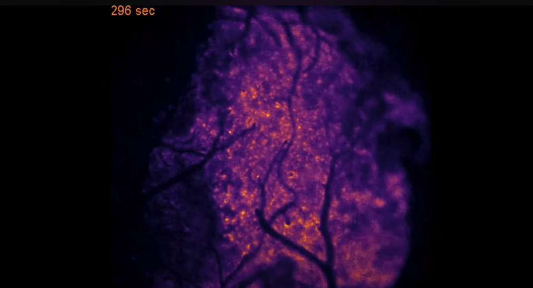
The enormous amounts of energy required by the human brain are nearly entirely produced by an oxygen-dependent kind of metabolism. Although it is well established that timely and effective oxygen supply is essential for normal brain function, scientists still don’t fully understand the exact mechanisms involved.
The flow of oxygen in mice’s brains has been captured in incredibly precise and visually arresting photos thanks to a novel bioluminescence imaging method that was published in the journal Science.
Researchers will be able to more thoroughly examine types of brain hypoxia, such as the deprivation of oxygen to the brain that happens during a stroke or heart attack, thanks to this easily replicable technology. The new study tool is beginning to shed light on the reasons why living a sedentary lifestyle may make illnesses like Alzheimer’s more likely to occur.
This research demonstrates that we can monitor changes in oxygen concentration continuously and in a wide area of the brain.
This provides us a with a more detailed picture of what is occurring in the brain in real time, allowing us to identify previously undetected areas of temporary hypoxia, which reflect changes in blood flow that can trigger neurological deficits.
Maiken Nedergaard
Luminescent proteins chemical relatives of the bioluminescent proteins found in fireflies are used in the new technique. These proteins, which have been applied to cancer research, work with a virus that instructs cells on how to make an enzyme that glows when exposed to light. Light is produced via the chemical interaction between the enzyme and furimazine, a second chemical molecule.
Similar to other significant scientific breakthroughs, the utilisation of this technique to see oxygen within the brain was fortuitously discovered. Originally, the luminous protein was going to be used to gauge brain calcium activity, according to Felix Beinlich, PhD, an assistant professor in the CTN at the University of Copenhagen. The investigation was delayed for several months until it was discovered that a mistake had occurred during the manufacturing of the proteins.
Beinlich decided to proceed with testing and refining the monitoring systems while he awaited a fresh supply from the manufacturer. The substrate was introduced into the brain by a craniotomy, and the virus was utilised to transfer instructions for the production of enzymes to astrocytes ubiquitous support cells in the brain that preserve the health and signalling capacities of neurons. The recordings showed activity, which was indicated by a bioluminescence that fluctuated in intensity. The researchers hypothesised that this activity represented the existence and concentration of oxygen, and they would subsequently confirm this.
The chemical reaction in this instance was oxygen dependent, so when there is the enzyme, the substrate, and oxygen, the system starts to glow.
Felix Beinlich
Also Read| New drugs for antibiotic-resistant bacteria, developed using generative AI
The researchers were able to see a sizable portion of the mice’s cortex in real-time, whereas the oxygen monitoring methods now in use only give information on a relatively tiny portion of the brain. By adjusting the oxygen content of the air the animals were breathing, the researchers were able to show that the bioluminescence’s intensity and oxygen concentration coincided. Moreover, variations in light intensity correlated with changes in sensory perception. For instance, the researchers observed the relevant area of the brain light up when the mice’s whiskers were stimulated with a blast of air.
This oxygen-dependent brain is oxygen-dependant, as seen by the rapid neurological damage that follows a heart attack or stroke. However, what occurs when tiny, momentary oxygen deprivations are applied to areas of the brain? Researchers had not even considered asking this issue until the Nedergaard lab team started attentively examining the fresh recordings. The little regions of the brain that were being monitored by the researchers would occasionally go dark for minutes at a time, indicating that the oxygen supply was being cut off.
A huge network of arteries and tiny capillaries, or microvessels, that penetrate brain tissue carry oxygen throughout the brain. After conducting several tests, the researchers concluded that capillary stalling a condition in which white blood cells momentarily obstruct microvessels and impede the transit of red blood cells that deliver oxygen was the cause of the oxygen deprivation. These regions, which the researchers dubbed “hypoxic pockets,” were more common in mice’s brains while they were at rest than when they were moving around. Capillary stalling has been seen in Alzheimer’s disease models and is thought to worsen with age.
The door is now open to study a range of diseases associated with hypoxia in the brain, including Alzheimer’s, vascular dementia, and long COVID, and how a sedentary lifestyle, aging, hypertension, and other factors contribute to these diseases.
It also provides a tool to test different drugs and types of exercise that improve vascular health and slow down the road to dementia.
Maiken Nedergaard
Source: University of Rochester Medical Center Newsroom
Journal Reference: M. Beinlich, Felix R., et al. “Oxygen Imaging of Hypoxic Pockets in the Mouse Cerebral Cortex.” Science, 2024, https://doi.org/adn1011.


