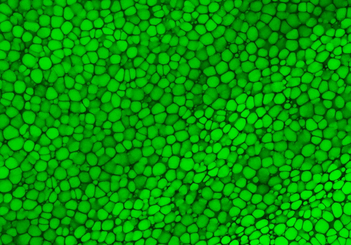
A novel kind of skeletal tissue has been found by an international research team headed by the University of California, Irvine. This discovery has enormous potential to advance tissue engineering and regenerative medicine.
The majority of cartilage is strengthened by an external extracellular matrix, but “lipocartilage,” which is found in mammals’ ears, noses, and throats, is special because it is packed with fat-filled cells called “lipochondrocytes” that give the tissue extremely stable internal support, keeping it soft and springy like bubbled packaging material.
The work explains how lipocartilage cells form and preserve their own lipid reservoirs while maintaining a consistent size. It was published online today in the journal Science. Lipochondrocytes, in contrast to regular adipocyte fat cells, do not contract or enlarge in reaction to the availability of food.
Lipocartilage’s resilience and stability provide a compliant, elastic quality that’s perfect for flexible body parts such as earlobes or the tip of the nose, opening exciting possibilities in regenerative medicine and tissue engineering, particularly for facial defects or injuries,
Currently, cartilage reconstruction often requires harvesting tissue from the patient’s rib – a painful and invasive procedure. In the future, patient-specific lipochondrocytes could be derived from stem cells, purified and used to manufacture living cartilage tailored to individual needs. With the help of 3D printing, these engineered tissues could be shaped to fit precisely, offering new solutions for treating birth defects, trauma and various cartilage diseases.
Maksim Plikus
When Dr. Franz Leydig discovered fat droplets in the cartilage of rat ears in 1854, he made the first discovery of lipochondrocytes. This discovery has since been mostly forgotten. Researchers from UC Irvine thoroughly described the molecular biology, metabolism, and structural function of lipocartilage in skeletal tissues using cutting-edge imaging techniques and contemporary biochemical tools.
They also discovered the genetic mechanism that essentially locks down the lipid stores in lipochondrocytes by inhibiting the action of enzymes that break down lipids and lowering the absorption of new fat molecules. The lipocartilage becomes brittle and stiff when its lipids are removed, underscoring the role that its fat-filled cells play in preserving the tissue’s ability to be both flexible and durable. Furthermore, the study observed that lipochondrocytes in certain species, like bats, form complex structures, such as parallel ridges in their large ears, which may improve hearing acuity by modifying sound waves.
Also Read: A Novel AI Predict Inner Cell Functions
The discovery of the unique lipid biology of lipocartilage challenges long-standing assumptions in biomechanics and opens doors to countless research opportunities,
Future directions include gaining an understanding of how lipochondrocytes maintain their stability over time and the molecular programs that govern their form and function, as well as insights into the mechanisms of cellular aging. Our findings underscore the versatility of lipids beyond metabolism and suggest new ways to harness their properties in tissue engineering and medicine.
Raul Ramos
Source: UC Irvine News
Journal Reference: Ramos, Raul, et al. “Superstable Lipid Vacuoles Endow Cartilage with Its Shape and Biomechanics.” Science, 2025, DOI: 10.1126/science.ads9960.
Last Modified:


