
 |
| Image: newcomersupply.com |
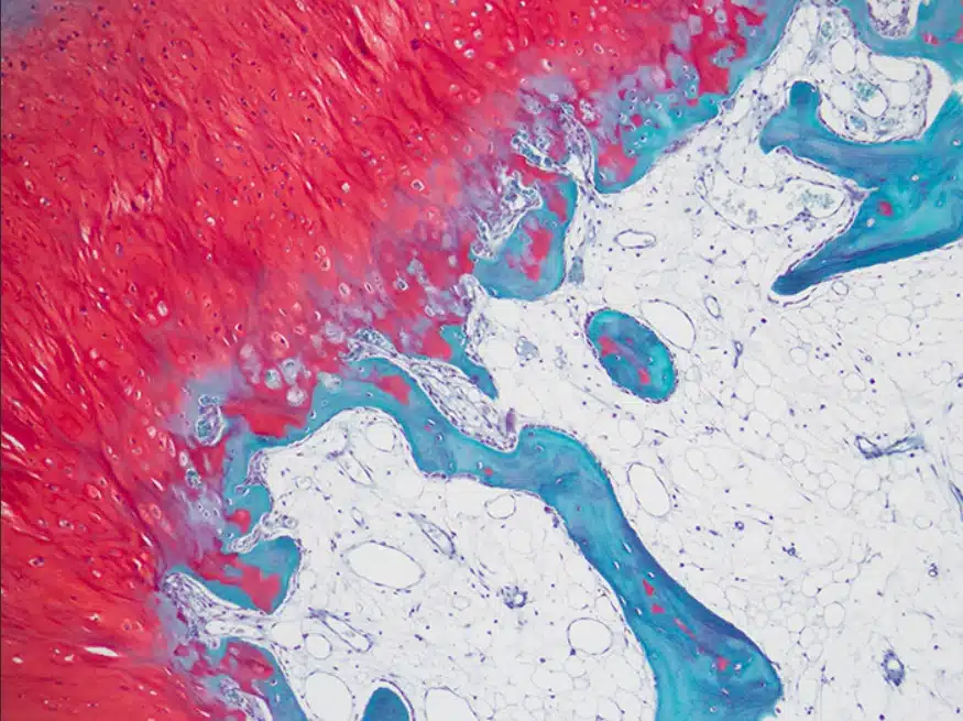
Safranin staining is a widely used technique in the field of biology and histology that allows researchers to visualize and study various biological structures with enhanced contrast. This staining method utilizes safranin, a red dye derived from saffron, which selectively binds to specific cellular components, providing researchers with valuable insights into cellular morphology, organization, and function. In this article, we delve into the principle behind safranin staining, its applications in different fields of research, and the benefits it offers in advancing our understanding of biological systems.

Principle of Safranin Staining
The safranin staining technique is based on the principle of selective affinity, where the safranin dye preferentially binds to specific cellular components, highlighting their presence within a sample. Safranin is known to exhibit a high affinity for nucleic acids, particularly DNA and RNA, making it an excellent tool for visualizing cell nuclei and other nucleic acid-rich structures. Additionally, safranin staining can also reveal cytoplasmic components, connective tissues, and certain organelles within cells.
Procedure for Safranin Staining
The process of safranin staining typically involves a series of steps, including sample preparation, staining, and visualization. Here is a generalized protocol for safranin staining:
- Fixation: The biological sample, such as tissue sections or cultured cells, is first fixed using an appropriate fixative, such as formalin or Paraformaldehyde, to preserve its structure and prevent degradation.
- Dehydration: The fixed sample is dehydrated using a series of alcohol washes, usually starting from lower concentrations (e.g., 70% ethanol) and gradually increasing to higher concentrations (e.g., 100% ethanol). Dehydration ensures proper penetration of the safranin dye into the tissue.
- Staining: The dehydrated sample is then immersed in a safranin solution, typically prepared by dissolving safranin dye in a suitable solvent such as water or ethanol. The duration of staining varies depending on the specific application and the desired intensity of staining.
- Differentiation: Excess safranin dye is removed by subjecting the stained sample to a differentiation step. This process typically involves rinsing the sample in a differentiating agent, such as acid alcohol, to remove excess dye from non-specifically stained regions.
- Dehydration and Mounting: The sample is rehydrated by passing it through a series of alcohol washes in reverse order, followed by clearing agents to remove any remaining alcohol. Finally, the sample is mounted on a slide using a mounting medium, such as a resin or glycerol, to preserve its structure and enhance the visibility of stained structures.
Applications of Safranin Staining
Safranin staining finds extensive applications in various fields of research, including:
- Histology: Safranin staining is widely employed in histopathology to visualize and differentiate different cell types, identify cellular structures, and study tissue architecture. It enables researchers to observe cellular details, such as nuclei, cytoplasmic components, and connective tissues, aiding in the diagnosis and study of diseases.
- Cytology: Safranin staining is valuable in cytological studies for highlighting cellular structures and components. It is commonly used in the examination of cell cultures and smear preparations, enabling the identification of different cell types, detecting abnormal cells, and studying cellular morphology.
- Botany: Safranin staining is extensively used in plant research to visualize various plant tissues and structures, such as plant cell walls, nuclei, and pollen grains. It aids in studying plant anatomy, growth patterns, and reproductive structures.
- Microbiology: Safranin staining is employed in microbiology to differentiate and visualize bacterial cells. It is commonly used in Gram staining, where safranin is used as a counterstain to color Gram-negative bacteria after the initial crystal violet staining.
Advantages of Safranin Staining
Safranin staining offers several advantages in biological research:
- Versatility: Safranin staining can be adapted to visualize a wide range of cellular structures and components, making it a versatile technique in various fields of biology.
- Selectivity: Safranin selectively binds to nucleic acids, allowing researchers to specifically target and visualize DNA and RNA within cells. This feature is particularly useful in distinguishing different cell types and studying cellular processes involving nucleic acids.
- Enhanced Contrast: Safranin staining enhances the contrast between stained structures and the surrounding background, improving the visibility and resolution of cellular details.
- Cost-effective: Safranin is a relatively inexpensive dye, making it an accessible staining method for researchers with limited resources.
Safranin staining is a valuable technique in biology and histology that facilitates the visualization and study of various cellular structures. Its selectivity, versatility, and cost-effectiveness make it a widely employed method in different research areas. By providing enhanced contrast and highlighting specific components within cells and tissues, safranin staining contributes significantly to our understanding of biological systems, aiding in disease diagnosis, cellular characterization, and advancements in various scientific disciplines.


![Good Laboratory Practice (GLP) – Overview [PDF]](https://cdn.biotechreality.com/wp-content/uploads/2023/10/GLP_Cover-min-300x169.png)