
The majority of research labs will be equipped with several centrifuge types, each with the ability to operate with different rotors. Small tabletop centrifuges can be used to gather DNA strands during ethanol precipitation or to pellet cells. Using an ultracentrifuge, one can separate distinct replicating DNA structures in a sucrose gradient or band plasmid DNA in a caesium chloride gradient.
Definition
Centrifugation is a technique used to separate cells, filter viral particles, precipitate DNA, and detect minute variations in molecular conformation. It is based on the molecular size, shape, and density of the product and is achieved by centrifugal force. Centrifugation is one of the most significant and often used methods in molecular biology labs.
Protein or DNA molecules in suspension are forced away from the center of rotation during centrifugation. Revolutions per minute, or rpm, is a unit of measurement used to express the speed at which this occurs. Relative centrifugal force (RCF), also known as the g force, is the force produced by the rotating rotor and is related to the square of the rotor speed and the radial distance (the distance the molecule is from the rotational axis).
Centrifugation is a technique used for the separation of particles using a centrifugal field. It causes denser particles to settle to the bottom of tubes, while low-density substances rise to the top. In this process, denser component of the mixture migrates away from the axis and lighter components migrate toward the axis. The sedimentation principle the rate or velocity at which sediment is proportionate to applied force underlies the operation of centrifuges, so as for the particles to settle down more quickly when a force is applied, that is greater than the earth’s gravitational pull. Usually, the particles are suspended in a particular liquid medium contained in tubes inside a rotor, which is positioned in the center of the drive shaft. The rate at which particles settle is directly correlated with the applied centrifugal force. Two main, different phases arise during centrifugation. In the vessel, they are Sediment, and in centrifugate, it is a supernatant liquid.

Centrifuge
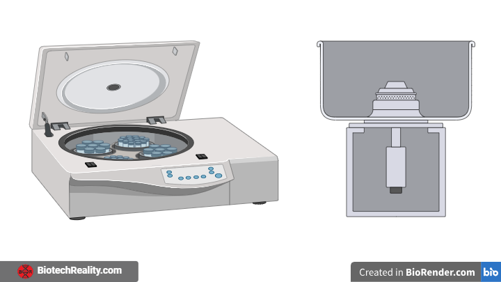
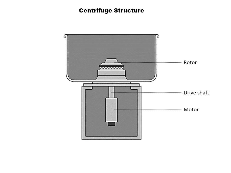
A centrifuge is used to perform the centrifugation process. This machinery is used to purify cells, viruses, proteins, nucleic acids, and subcellular organelles. It is usually found in most labs, including academic, clinical, and research institutions. The whole machine is capable of regulating temperature and speed to facilitate the separation process. The structure of the centrifuge has the following features.
Centrifuge has three basic components:
- Rotor
- Driveshaft
- Motor
The tubes or bottles containing the liquids to be centrifuged are held in place by the rotor. On the drive shaft, rotors of various sizes and types can be attached. The motor, which powers the rotor and allows the tube to spin at an assigned speed, is attached to the driving shaft. A centrifugal force is applied to the suspension of particles when the rotor rotates around its central axis.
Centrifuge diagram
The centrifugal force exerted on the suspended particles is counteracted by two forces. They are the forces of buoyancy and friction. Particles sediment when their rotational force, known as centrifugal force, is greater than the opposing forces of buoyancy and friction.
The rate of sedimentation depends on:
- The applied centrifugal field
- Size, density, and radius of the particle
- Density and viscosity of the suspending medium
The rate of centrifugation is expressed as revolutions per minute (RPM), which is specified by the angular velocity (ω).
One revolution per minute (rpm) = (ω×60)/2π
ω = (2π×rpm)/60, Where ω= angular velocity (radians/second).
Types of rotors
Rotors come in three different types. These are swinging bucket rotors, vertical tube rotors, and fixed angle rotors.
- Fixed angle rotors: The tubes are positioned between 14 and 40 degrees from the vertical axis. Particles radially travel a short distance. For differential centrifugation, it is helpful. In this case, the tube reorients as the rotor accelerates and decelerates.
- Vertical tube rotors: They are kept vertically in line with the rotor axis. The particles move a short distance and split apart faster. The drawback is that after centrifugation, the pellet could return to solution.
- Swinging bucket rotors: The rotor swings out to the horizontal position when the rotor accelerates. The particle travels for a longer distance and thus it may allow better separation.
Types of Centrifuges
1. Low-speed centrifuge
RCF (Relative Centrifugal Force) values can reach 6000 g, and the centrifugation speed ranges from 1 to 6000 rpm. Without a way to regulate the sample’s temperature, these instruments typically run at room temperature. It is possible to employ a swinging bucket and fixed-angle rotors. Low-speed centrifuges are especially useful for the rapid sedimentation of coarse precipitates or red blood cells as pellets at the bottom of the tube.
2. High-speed centrifuge
High-speed centrifuges are traditional centrifuges used in laboratories and are widely used in biological, pharmaceutical, and other scientific research, universities, and production sectors. It uses high-speed rotation to create centrifugal force to separate liquids and solids or liquids mixed with solids. They are suitable for rapid separation of samples. The centrifuges have a maximum speed of 15,000 to 20,000 rpm and they are useful for collecting microorganisms, larger cell organelles, and protein precipitated by ammonium sulfate. These types of centrifuges use three types of rotors: angled rotors, horizontal rotors, and vertical rotors. High-speed centrifuges are equipped with machinery to control the speed and temperature required to analyze sensitive biomolecules.
3. Small bench-top centrifuge
Small bench-top centrifuges are the simplest and least expensive and are often used in laboratories for the sedimentation of blood samples and urine, and body fluid separation. It is driven by an electric motor that rotates the tube around a fixed axis and creates a force perpendicular to the tube. The maximum speed range is up to 4000 – 6000 rpm. A small bench-top centrifuge has a rotor with a standard tube holder. The flow of air around the rotor controls the rotor temperature. These centrifuges exist in different types and designs.
4. Microcentrifuge
These centrifuges are used to separate small volumes (0.5 to 2 µl) of samples. Microcentrifuge typically operates at approximately 12,000-13,000 rpm. It is used for the separation of cell organelles like nuclei and DNA. Microcentrifuges, also called microfuges, use sample tubes that are smaller in size when compared to the standard test tubes used in larger centrifuges. Some Microcentrifuges come with adapters to facilitate the use of large and small tubes.
5. Continuous flow centrifuge
Continuous flow centrifuges are rapid centrifuges that can centrifuge a large volume of the sample without affecting the sedimentation rate. It is used for the isolation of bacteria cells from large volumes of culture media. They have a Shorter path length that facilitates the precipitation of residues from the supernatant, thus maintaining the rate of the process. They also have a larger capacity, which saves time because there is no need to load and unload samples as required in conventional centrifuges. This centrifuge can spin up to 1 litre of sample in 4 hours or less.
6. Ultracentrifuge
An ultracentrifuge is a centrifuge that operates at very high speeds to separate small molecules such as ribosomes, proteins, and bacteria. These types of centrifuges have a cooling system that helps to balance the heat generated by the spinning power. These centrifuges can operate at speeds up to 150,000 rpm. In addition to separation, ultracentrifuges can also be used to determine properties of macromolecules such as size, shape, and density. There are two types of ultracentrifuge, preparative and analytical ultracentrifuge. In preparative ultracentrifuge maximum speed range up to 80,000. The rotor chamber is refrigerated and sealed. They are fitted with a flexible drive shaft system which minimizes vibration caused by slight rotor imbalance that arises due to unequal loading of the centrifuge tube. In analytical ultracentrifuge, maximum speed ranges up to 70,000. 3 types of Optical system is available to evaluate the sedimenting material. They are a light absorption system, an Alternative schlieren system, and a Rayleigh interferometer system.
7. Gas centrifuge
Gas centrifuges are specifically designed to separate gases based on isotopes and are mainly used for the extraction of uranium. This centrifuge is based on the same principle of centrifugal force as all other centrifuges in which molecules are separated according to their size. These centrifuges are arranged in stages so the gas is separated by two units based on isotopes and then transferred to the next centrifuge for processing.
8. Vacuum centrifuge
Vacuum centrifuges are used in chemical and biological laboratories for the effective evaporation of solvent from the sample and use centrifugal force, vacuum, and heat to accelerate the process. These centrifuges can process large amounts of samples. Rotary evaporators are used to remove unwanted solvents. The centrifuge works by reducing the chamber pressure, which lowers the temperature of the sample.
9. Refrigerated centrifuge
Refrigerated centrifuge with a temperature range of -20°C to -30°C. The centrifuge is available in different models and features a temperature control system, which is important for many purposes that require lower temperatures as well as a rotor and sample tube holder. These centrifuges have a speed range of up to 60,000 and are ideal for separating a variety of biomolecules. These are also used to collect rapidly dividing cells such as yeast, chloroplasts, and red blood cells.
Also Read


Centrifugation technique
Depending on the purpose of use, centrifugation can be divided into two types: Preparative and analytical centrifugation. (Preparative is just for a preparative scale separation and in analytical centrifugation, analysis of certain parameters is performed.)
Preparative centrifugation technique
It focuses on the actual isolation, purification, and separation of viruses, polysomes, lipoproteins, ribosomes, plasma membranes, and nucleic acids. Following centrifugation, the rotor is to be stopped, and each tube’s gradient is gradually fed through to isolate each individual component. Following each run, there is no visual readout available for examination. Molecular and organelle separation based on sedimentation rate.
Types of preparative centrifugation:
Differential centrifugation
This technique, which is based on variations in the sedimentation rates of particles of varying sizes and densities, is employed to isolate certain organelles. By increasing the applied centrifugal field, the material to be separated is split centrifugally into a number of fractions. Every stage’s centrifugal field is selected to allow a certain kind of material to settle within the predefined centrifugation period, producing a pellet of particles and supernatant. A single centrifugation cannot produce a pellet of the heaviest particle. The majority of material that sediments slowly stays in the supernatant. In the buffer, the sample solution was homogenized. After that, the sample is put into a centrifuge tube and operated for a certain amount of time at a predetermined temperature and centrifugal force. A precipitate that separates from the supernatant will form at the tube’s bottom at the conclusion of this operation. After adding the supernatant to a fresh centrifuge tube, centrifuge for the designated amount of time and temperature at a different speed. The pellet and supernatant were separated. Until all components are separated, keep going through this procedure.
Density gradient centrifugation
Centrifugation is a technique used to separate biological particles of similar sizes but different densities. The basis for the separation is the molecule’s density as it moves across the density gradient while being driven by centrifugal force. This separation method makes use of a variety of media, such as silica, polysaccharides, and inorganic salts. This method uses a material having a gradient in density, where the density has to be raised or lowered. A centrifugal force is produced as the sample spins due to the molecules in the sample moving through the medium. Denser molecules start to descend as they pass over the density gradient. At a certain point, the density of the particles matches that of the surrounding medium, and the molecules become suspended. There are two types of density gradient centrifugation, rate zonal technique and isopycnic technique.
Rate zonal technique
This method is applied to the separation of hormones, ribosomal subunits, and enzymes. Particle size, shape, and density differences, medium viscosity and density, and the applied centrifugal field all affect separation. Using this method, the sample solution must be properly positioned at the top of the liquid density gradient. whose greatest density is not more than the densest particles that need to be separated. The purpose of the gradient is to provide a viscosity gradient that aids in improving the gradient’s resolution and stable the liquid column in the tube against movement brought on by conventional current. After that, the sample is centrifuged for a long enough period of time to enable the particles to cross the gradient and form a distinct zone.
Isopycnic centrifugation
This method is independent of time and relies on the buoyant density of the particle rather than its size or form. It can separate subcellular organelles but not soluble proteins with the same density since it is designed to separate particles of identical sizes but different densities. On top of the density gradient is where the sample is positioned. Centrifugation causes sedimentation until the particle’s gradient and buoyant densities are equal. At the isodensity point, there is no further sedimentation. These methods involve the use of sucrose, silica, and a heavy metal salt (caesium). CsCl is combined with the sample. The CsCl molecule sediments when the CsCl solution is centrifuged, creating a concentration gradient and subsequently a density gradient. The sample molecules were evenly dispersed throughout the tube, and when they got to a place where the density of the solution matched their own buoyant density, they joined together to create zones.
Analytical centrifugation
It is employed to separate pure macromolecules, with the centrifugal force and density serving as the bases of separation. They use specifically made rotors and detectors to continually monitor the sedimentation process of materials, allowing for the study of the relative molecular mass and form of the substance with minimal material requirements. Three optical systems fluorescence, reflectance, and absorbance are included in analytical ultracentrifuges to provide quick, accurate, and focused sedimentation examination. A little sample (20–120 mm3) should be placed in analytical cells and then into an ultracentrifuge. After turning on the ultracentrifuge, the randomly dispersed biomolecules begin to move radially outward from the center of rotation due to centrifugal force.
Author: Athulya B S | Edited: Ajmal Aseem
Last updated: