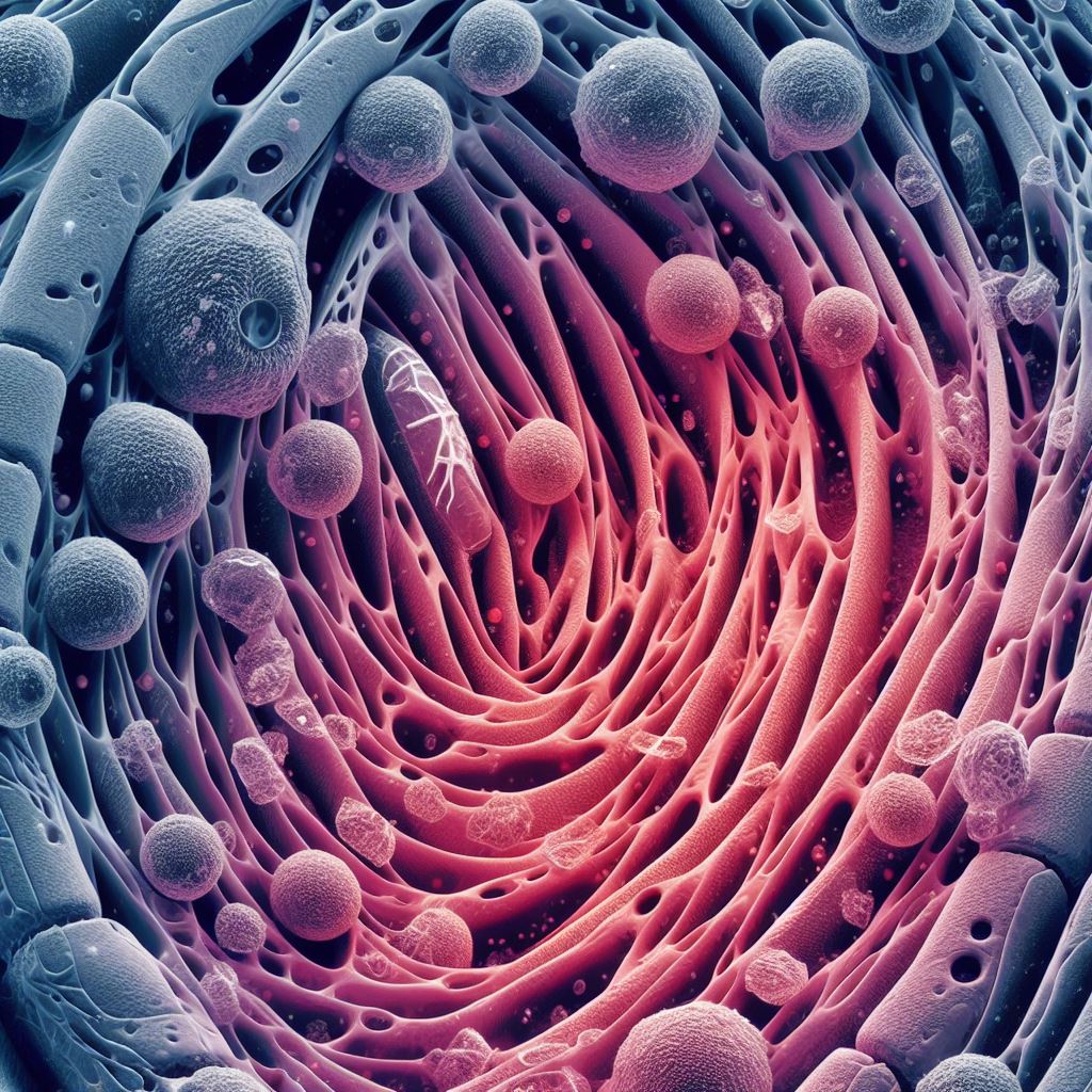
To restore lost cardiac function, regenerative heart treatments entail grafting cardiac muscle cells into injured cardiac regions. However, there is a substantial documented risk of arrhythmias after this surgery. In a recent study, Japanese researchers explored a unique technique that includes directly injecting human stem cell-cultured “cardiac spheroids” into injured ventricles. The promising results in monkey models demonstrate the potential of this approach.
Cardiovascular illnesses continue to rank among the leading causes of mortality globally, with industrialised nations having higher rates of this condition. Heart attacks sometimes referred to as myocardial infarctions, are becoming more frequent and cause a considerable number of fatalities annually.
Millions of cardiac muscle cells are usually destroyed during a heart attack, weakening the heart. For individuals experiencing (or expected to have) heart failure, heart transplants are now the only therapeutically feasible alternative because humans are unable to grow cardiac muscle cells on their own. Understandably, the medical world is quite interested in alternative medicines given that complete heart transplants are costly and donors are hard to come by.
Human induced pluripotent stem cells (HiPSCs) represent one exciting approach that is gradually gaining popularity in the field of regenerative cardiac treatment. In a nutshell, hiPSCs are cells that are produced from adult cells and can be “reprogrammed” into a whole new cell type, such as cardiomyocytes, which are the cells that make up the heart. Some lost functioning of the heart may be regained by transplanting or injecting cardiomyocytes produced from HiPSCs into damaged regions. Sadly, research has shown that using this strategy may raise the possibility of arrhythmias, which would be quite problematic for clinical trials.
A Japanese research team from Shinshu University and Keio University School of Medicine has attempted injecting “cardiac spheroids,” which are produced from HiPSCs, into myocardial infarcted animals as a novel approach to regenerative heart treatment. The lead author of this work, Professor Yuji Shiba of Shinshu University’s Department of Regenerative Science and Medicine, published it in the journal Circulation.
The first author, Hideki Kobayashi, as well as Koichiro Kuwahara and Shugo Tohyama from Shinshu University School of Medicine’s Department of Cardiovascular Medicine and Keiichi Fukuda from Keio University School of Medicine’s Department of Cardiology, were among the team members.
The researchers used a unique technique in which they cultured HiPSCs in a medium that caused them to differentiate into cardiomyocytes. Following a meticulous extraction and purification process, the researchers injected around 6 × 10⁷ cardiac spheroids three-dimensional clusters of cardiac cells into the injured hearts of macaques that eat crabs (Macaca fascicularis). Throughout twelve weeks, they kept an eye on the animals’ health and measured their heart rate regularly. After that, they examined the tissue of the monkeys’ hearts to determine whether cardiac spheroids could repair the injured heart muscle.
Initially, the group confirmed that HiPSCs had been properly reprogrammed into cardiomyocytes. Through electrical measurements at the cellular level, they saw that the cultivated cells had potential patterns that are characteristic of ventricular cells. Also, the cells reacted to several well-known medications as predicted. The discovery that the cells highly expressed adhesion proteins including N-cadherin and connexin 43, which would facilitate their vascular integration into an existing heart, was the most significant finding.
Subsequently, the cells were sent 230 km to Shinshu University from Keio University’s production plant. After being stored at 4 °C in regular containers, the cardiac spheroids travelled for four hours without any issues. This implies that the cells might be transported to clinics without the need for drastic cryogenic procedures, which would lower the cost and facilitate the adoption of the suggested strategy.
Lastly, the injured heart ventricle of the animals was directly injected with cardiac spheroids or a placebo. Within the therapy group, just two patients experienced transitory tachycardia (rapid pulse) in the first two weeks of the monitoring period, according to the researchers, indicating that arrhythmias were extremely rare. By using computed tomography and echocardiography, the researchers were able to verify that, at four weeks, the hearts of the treated monkeys were able to pump more blood than the hearts of the control group, as evidenced by improved left ventricular ejection fraction.
The cardiac grafts were mature and correctly attached to pre-existing existent tissue, as shown by histological investigation, which confirmed the findings of earlier studies.
Also Read| Researchers created artificial cells that mimic the original cells
HiPSC-derived cardiac spheroids could potentially serve as an optimal form of cardiomyocyte products for heart regeneration, given their straightforward generation process and effectiveness.
We believe that the results of this research will help solve the major issue of ventricular arrhythmia that occurs after cell transplantation and will greatly accelerate the realization of cardiac regenerative therapy.
Assistant Professor Kobayashi
Source: Shinsu University Topics
Journal Reference: Kobayashi, Hideki et al. “Regeneration of Nonhuman Primate Hearts With Human Induced Pluripotent Stem Cell-Derived Cardiac Spheroids.” Circulation, 10.1161/CIRCULATIONAHA.123.064876. 26 Apr. 2024, doi:10.1161/CIRCULATIONAHA.123.064876
Last Modified:


